


 Foundational characteristics of cancer include proliferation, angiogenesis, migration, evasion of apoptosis, and cellular immortality. Find key markers for these cellular processes and antibodies to detect them.
 The SUMOplot™ Analysis Program predicts and scores sumoylation sites in your protein. SUMOylation is a post-translational modification involved in various cellular processes, such as nuclear-cytosolic transport, transcriptional regulation, apoptosis, protein stability, response to stress, and progression through the cell cycle.
 The Autophagy Receptor Motif Plotter predicts and scores autophagy receptor binding sites in your protein. Identifying proteins connected to this pathway is critical to understanding the role of autophagy in physiological as well as pathological processes such as development, differentiation, neurodegenerative diseases, stress, infection, and cancer.
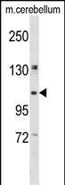
KIF5B Antibody (C-term)
Affinity Purified Rabbit Polyclonal Antibody (Pab)
- SPECIFICATION
- CITATIONS
- PROTOCOLS
- BACKGROUND

Application
| WB, E |
|---|---|
| Primary Accession | P33176 |
| Other Accession | Q2PQA9, Q61768, NP_004512.1 |
| Reactivity | Human, Mouse |
| Predicted | Rat |
| Host | Rabbit |
| Clonality | Polyclonal |
| Isotype | Rabbit IgG |
| Calculated MW | 109685 Da |
| Antigen Region | 867-895 aa |
| Gene ID | 3799 |
|---|---|
| Other Names | Kinesin-1 heavy chain, Conventional kinesin heavy chain, Ubiquitous kinesin heavy chain, UKHC, KIF5B, KNS, KNS1 |
| Target/Specificity | This KIF5B antibody is generated from rabbits immunized with a KLH conjugated synthetic peptide between 867-895 amino acids from the C-terminal region of human KIF5B. |
| Dilution | WB~~1:1000 E~~Use at an assay dependent concentration. |
| Format | Purified polyclonal antibody supplied in PBS with 0.09% (W/V) sodium azide. This antibody is purified through a protein A column, followed by peptide affinity purification. |
| Storage | Maintain refrigerated at 2-8°C for up to 2 weeks. For long term storage store at -20°C in small aliquots to prevent freeze-thaw cycles. |
| Precautions | KIF5B Antibody (C-term) is for research use only and not for use in diagnostic or therapeutic procedures. |
| Name | KIF5B (HGNC:6324) |
|---|---|
| Synonyms | KNS, KNS1 |
| Function | Microtubule-dependent motor required for normal distribution of mitochondria and lysosomes. Can induce formation of neurite-like membrane protrusions in non-neuronal cells in a ZFYVE27-dependent manner (By similarity). Regulates centrosome and nuclear positioning during mitotic entry. During the G2 phase of the cell cycle in a BICD2- dependent manner, antagonizes dynein function and drives the separation of nuclei and centrosomes (PubMed:20386726). Required for anterograde axonal transportation of MAPK8IP3/JIP3 which is essential for MAPK8IP3/JIP3 function in axon elongation (By similarity). Through binding with PLEKHM2 and ARL8B, directs lysosome movement toward microtubule plus ends (Probable). Involved in NK cell-mediated cytotoxicity. Drives the polarization of cytolytic granules and microtubule-organizing centers (MTOCs) toward the immune synapse between effector NK lymphocytes and target cells (PubMed:24088571). |
| Cellular Location | Cytoplasm, cytoskeleton {ECO:0000250|UniProtKB:Q2PQA9}. Cytolytic granule membrane. Lysosome membrane; Peripheral membrane protein; Cytoplasmic side Note=Uniformly distributed between soma and neurites in hippocampal neurons. {ECO:0000250|UniProtKB:Q2PQA9} |

Thousands of laboratories across the world have published research that depended on the performance of antibodies from Abcepta to advance their research. Check out links to articles that cite our products in major peer-reviewed journals, organized by research category.
info@abcepta.com, and receive a free "I Love Antibodies" mug.

Provided below are standard protocols that you may find useful for product applications.
Background
Microtubule-dependent motor required for normal distribution of mitochondria and lysosomes (By similarity).
References
Seeger, M.A., et al. J. Biol. Chem. 285(11):8155-8162(2010)
Trejo, H.E., et al. FASEB J. 24(2):374-382(2010)
Morton, A.M., et al. Biochem. Biophys. Res. Commun. 391(1):388-393(2010)
Zadeh, A.D., et al. J. Physiol. (Lond.) 587 (PT 19), 4565-4574 (2009) :
Wong, Y.L., et al. Biophys. J. 96(7):2799-2807(2009)
If you have used an Abcepta product and would like to share how it has performed, please click on the "Submit Review" button and provide the requested information. Our staff will examine and post your review and contact you if needed.
If you have any additional inquiries please email technical services at tech@abcepta.com.
Ordering Information
Other Products
Shipping Information
